When the heart goes out of rhythm: Treating Atrial Fibrillation – Part III

Why are we so concerned about Atrial Fibrillation (A Fib)? Although the rapid heart rate associated with untreated A Fib can produce symptoms on its own, it can also precipitate or aggravate heart failure and worsen symptoms of angina from myocardial ischemia. However, the major cause for concern is its propensity to produce clots inside the cardiac chambers, mainly the left atrium. Even in patients with transient A Fib, the blood can pool in the atrial chamber leading to clots that could break loose and travel to peripheral organs, especially the brain producing a stroke.
The first dictum in the treatment of any disease is to treat the cause. If hypertension or coronary artery disease is the problem, then appropriate management with BP-lowering medicines, angioplasty or coronary bypass surgery and lifestyle changes like stopping smoking are all indicated. If it is related to an excess of thyroid hormone circulating in the blood, then anti-thyroid medications or even surgery for the thyroid may be necessary.
The two aspects in the treatment are: 1. Control the heart rate and rhythm. If the heart rate is faster than normal, slow it down through a variety of medicines like beta blockers, calcium channel blockers or even the old-fashioned digoxin; this is ‘rate control.’ 2. Restore the fibrillation back to regular sinus rhythm – called rhythm control. Many powerful drugs like amiodarone, dofetilide and sotalol are available for this purpose. These are potent drugs with side effects and hence you need to make a special effort to learn about them. Sometimes, drugs may not do the job; so electrical cardioversion is done by giving a gentle electric shock to the chest wall under controlled conditions by the cardiologist, which often work well. For the average person with A Fib, large studies have found that rhythm control and rate control work equally well. However, if rhythm can be kept normal always, there is less chance for clot formation and embolism.
If the patient suffers from recurrent episodes of A Fib, there are two invasive approaches to restore sinus rhythm. One is using radiofrequency waves via a special catheter inserted into the heart. The tip of this device that emits radiofrequency waves will zap the focus or foci causing A Fib. The other is ‘maze operation,’ an open heart surgery that creates scars in both atrial walls aimed at disrupting the path of abnormal impulses, which in turn restores regular sinus rhythm.
While restoration of normal rhythm may be the main objective of treatment of A Fib, the most dreadful consequence of the disease is stroke from cerebral embolism. A clot in the left atrium, especially the left atrial appendage, the commonest region for clot formation, is the main source for such embolic episodes. A Fib is responsible for 20 percent of all strokes and may be the commonest cause in older patients who do not suffer from mitral valvular disease. Hence, blood thinning medicines like warfarin are an important part of the preventive treatment. Three out of four A Fib related strokes can be prevented with proper management.
There is an assessment test called CHADS2 to determine your risk of stroke. One point is assigned for each of these conditions: congestive heart failure, hypertension, age 75 or older and, diabetes. Two points will be assigned if you had a previous stroke. The maximum score one can get is 6 points which is associated with an annual stroke rate of 18 percent! So, it’s critical for all patients with A Fib to understand the implications of the diagnosis and proper management of concomitant conditions. Needless to say, those who are on anticoagulants like warfarin or novel ones such as Xarelto, Pradaxa and Eliquis should know all aspects of these drugs and how to monitor them.
As mentioned before, more than 90 percent of the clots come from the left atrial appendage, a small ear-shaped pouch sticking out of the left atrial wall. This can be closed off by implanting a special device called Watchman device that prevents clots from migrating out. Another procedure is excision of left atrial appendage by surgical method. Both these treatments give the patient an anticoagulant free alternative to manage their A Fib but they may have to take antiplatelet agents like clopidogrel and aspirin. You must discuss with your doctor in detail and understand all the pros and cons of management of the condition. Also, pay special attention to maintain a healthy lifestyle.
* This concludes the mini-series on Atrial Fibrillation
M.P. Ravindra Nathan, M.D., is a cardiologist and Emeritus Editor of AAPI Journal. His book “Stories from My Heart” was recently released. (www.amazon.com or www.bn.com).
The Players Championship and Golfer’s eyes

With the recently concluded 43rd Players Championship at Sawgrass in Ponte Vedra – a flagship event of the PGA tour and tens of thousands of people from around the world involved – we can surely comprehend the universal impact of this once private sport.
A dream realized by Deane Beman, founding commissioner of the PGA tour who relentlessly propelled this magnificent sport to the world. When I recently concluded his eye surgery, he taught me that, “In golf, learning to putt should come before learning to drive.”
Golf has now become a sport of the elite and the commons and like all things in medicine, pathology then strikes both classes equally.
Golf is a sport that is visually demanding besides being technique and technology dependent. It also requires players to be out in the sun resulting in red eyes with tissue growths on the whites of the eyes commonly called Golfer’s eyes.
I like to classify vision demands used in golf into the following categories for ease of understanding and future surgical correction:
-
Quantity of vision like 20/40, 20/20, etc.
-
Quality of vision like:
a: Contrast sensitivity to decipher different levels of visual inputs in different lighting conditions;
b: Depth perception to best apply learned techniques in three dimension;
c: Spatial resolution to plan and direct putting and long drives effectively.
Finally, all the vision inputs and resolutions along with eye-hand coordination lead to perfect execution for what the mind has planned in this mentally intuitive sport.
This is true for novice as well as professional golfers alike.
Vision Errors
Golfers who may wear contact lenses or glasses may have nearsightedness (Myopia), farsightedness (Hyperopia), astigmatism (Football-shaped cornea). With technology today, we can diagnose and measure to the minutest level of vision involvement and thence correct using computer drive, micron-precision lasers as in NexGen Lasik techniques. Today, “no-blade” techniques allow for painless surgery with rapid healing and predictable outcomes.
As golfers advance in age (beyond 40 years), they need reading glasses because the natural lens inside the eye stops zooming (no more “tele” and “wide” zooming like a camera does) and hence they need to keep their score cards at further distance with their arms outstretched to read or record. This condition called Presbyopia can be addressed with new-generation laser techniques.
With further aging (early 60s), the vision gets cloudy and dim due to cataracts (a cataract is a cloudy transformation of our crystal clear, natural lens). This cataract then can be removed using modern laser techniques and replaced by an artificial lens implant that can in most people allow them to see without glasses at distance and near. Such lens implants can be monofocal, multifocal like ReStor, Rezoom, Tecnis or Symphony or accommodative like Crystalens.
Additionally, cataract surgery can be planned as two stages. Following cataract surgery, patients can undergo Lasik surgery to correct any residual astigmatism to thus completely fix the optics of the eyes for a glasses-free vision experience for life.
Even patients who have undergone previous cataract surgery and did not have a choice of these advanced, multifocal/progressive lens implants at that time can avail of new- generation Lasik surgery to help get rid of glasses altogether.
Golfer’s Eyes
Another condition rampant among golfers is pterygium, a yellow-red lesion on the white of the eye that can start as a cosmetic blemish and become an unsightly wedge-shaped growth. If left untreated, it may eventually cover the cornea of the eye and adversely affect vision. Pterygium is usually the result of exposure to sun, a professional hazard for golfers.
A lot of golfers have pterygiums and we usually counsel them to wear sunglasses with ultraviolet protection to prevent further damage, but when they are advanced, they require surgery. “No-Stitch” surgical technique using human placenta is the answer to this malady.
So, before heading out to golf as you apply your sunscreen, do remember to protect your eyes with sunglasses and a wide-brimmed hat, if possible. This may ultimately contribute to “Fashion on the Fairway.”
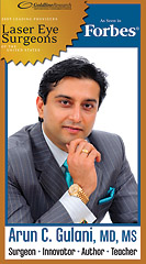
Arun C. Gulani, M.D., M.S., is director and chief surgeon of Gulani Vision Institute in Jacksonville. He can be reached at [email protected] or visit www.gulanivision.com